Wednesday, August 17, 2011

beta blockers in COPD

It has been a looong gap ....moving and starting a fellowship in a new place is a little tough. Anyway I have settled down in my new place now. 
Let me start with a short blog..regarding a discussion between a pulmonary fellow and the primary team that I happened to hear while doing my usual hyponatremia consult!
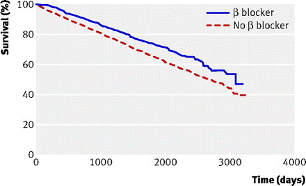
Traditionally betablockers were not used in patients with COPD or asthma due to bronchospam. But using a cardioselective agent may not afect the airways.But a recent retrospective study in bmj went a step ahead and showed that it may be even beneficial in patients with COPD. 
Of nearly 6000 patients with different stages of COPD ,they found that there was a 22% reduction in all cause mortality from beta blockers. The benifit was shown across all stages of COPD. With regards to lung function, they did not affect FEV1/FVC. also betablockers seemed to have additive benifits on hospital admissions. This benifit was seen independent of presence of cardiovascular disease.

Figure


A somewhat related study published by one of my senior resident(Int J Chron Obst Pulm Disease 2010)  showed that patients with COPD may have unrecognised myocardial injury. May be betablockers help with this and this is what we are seeing here. 
Time to change practice!!

Wednesday, May 25, 2011

Osteoporosis --- Screening & Treatment

Osteoporosis is very much underdiagnosed, and under treated! I recently did my practice based learning exercise on Osteoporosis, and sadly...I havent screened or treated many of my patients!!


So lets see who needs to be screened, and who needs treatement...
DEXA Scan and scoring  - Screening /diagnosing is done by DEXA scan . The 2 preferred sites of scanning are lumbar spine and proximal femur,as these places show the earliest sign of osteoporosis. So next time when you see a score based on a heel DEXA ....it is not a complete test! A T-score( representing the number of standard deviations between the pts bone mineral density and a healthy young adult of same sex) less than -2.5 is called osteoporosis, and a score between -1 to -2.5 is osteopenia. 
The Z-score is used to classify the type of osteoporosis. A score below 1.5 indicates primary osteoporosis, which is age related. A score of 1.5 and higher indicates secondary osteoporosis, which is associated with calcitonin imbalance, malabsorption conditions ,alcohol abuse, smoking, medications etc.


Screening - The following are the group of people who need DEXA for screening

  • women 65yrs and older,
  •  men 70 yrs and older,
  • Loss of height of >2 inches (before DEXA...do a vertebral fracture survey...and if +ve, treat as osteoporosis)
  • postmenopausal women or, men >50 yrs with risk for fracture including h/o hip fracture in parents, current smoker or alcoholic, rheumatoid arthritis.
  • chronic glucocorticoid therapy(5mg/d or more of prednisone for >3 mon)
  • pts with medical diseaseassociated with decreased BMD(eg. renal disease, hyperthyroisim, cushings etc)
  • pts on medications that might reduce BMD ( anticonvulsants, PPI,Cyclosporine, SSRI etc
Who needs treatment -  
  • Anyone who has sustained a fragility fracture of hip or spine
  • Those who have a T-score > or = 2.5, after evaluation for secondary causes of osteoporosis.
  • Those who have osteopenia(a score between -1 & -2.5) if their 10 year probablity of osteoporotic fracture is >20% or 10 yr risk of hip fracture >3%. ( the risk called FRAX ..can be calculated online)
Treatment -  Bisphosphonates are the mainstay of treatment. A quick note is...of all the treatment modalities, Ibandronate, Calcitonin, Raloxifene, and Teriparatide are the ones which have not shown any hip fracture reduction. All of them reduce the incidence of spine fracture though.

Tuesday, May 17, 2011

Pre exposure prophylaxis for HIV

A homosexual male who is an iv drug abuser walks into the office...asking ways to help him prevent contacting HIV..with his high risk behaviour!! Apart from advising on safe sex and risks of sharing needles(which are most important..and at the same time cannot be relied at all times due to non adherence)....do we have any medications to offer him??


Lets look at the iPREX study published in NEJM Dec 2010. This was a placebo-controlled, double-blind, randomized trial involving 2499 subjects(men who have sex with men) to see if a combination of Emtricitabine-Tenofivir(FTC-TDF) once daily reduced the incidence of HIV over a f/u period of 1.2 years. Of the 100 incident HIV infections during the study period, 64 occurred in the placebo group and 36 in the FTC–TDF group, with an efficacy of 44% and a 95% CI of 15 to 63. An important finding to note is that, in the FTC–TDF group, the study drug was pharmacologically detected in 51% of subjects who remained free of HIV infection but in only 9% of those who became infected. So exposure to FTC–TDF was associated with a reduction in HIV acquisition. Main issues in putting this into practice has a few concerns. Few of them are drug adherence(very poor in the study), long term outcome, side effects(quite a few in the study drug group had renal impairement which rverted to normal on discontinuation of drug!). All these issues have been nicely discussed in the letter following the study( another arrow in the Quiver.NEJM Dec 2010)
Now the trial has been extended to look for long term outcomes and ways to improve adherence. results are expected by 2013.(HIV & TB News)


An earlier study , CAPRISA 004 in which a total of 889 sexually active, HIV-uninfected women (age range, 18–40) received either 1% tenofovir gel or placebo gel, with instructions to administer the gel intravaginally within 12 hours before sex and also within 12 hours after sex. Mean follow-up was 18 months.The incidence of HIV was 5.6 /100 person-years in the tenofovir group versus 9.1 /100 person-years in the placebo group, for an incidence rate ratio (IRR) of 0.61 (95% CI, 0.40–0.94; P=0.017). Shows promise for pre exposure prophylaxis.


In this context ...FEM-PrEP trial has to be mentioned. This study evaluated if oral daily FTC-TDF prevented HIV in a group of high risk women. The study was stopped last month after an interim analysis didnt find any benefit compared to placebo.(AVAC April 2011). Full analysis of trial should be available shortly.(may be related to adherence issues!!)


VOICE trial - Vaginal and Oral Interventions to Control the Epidemic .........is an ongoing study on high risk women comparing FTC-TDF, tenofovir tablet alone , or tenofovir gel. This study might shed light into ..which of these will be more effective. By this time we will also have the long term safety and compliance outcomes on the i-PREX study.
May be 2013 might be a year the world would see a...  'HIV prevention pill'  !!!

Friday, May 13, 2011

The "aura" of migraine

Migraine can be with or without aura. What exactly comprises this aura. The prodromal  and associated  migraine symptoms may me mistaken as aura. Lets see what comprises the so called aura.  It is the migraine with aura that is related to an increased incidence of ischemic stroke, and is a contraindication for taking oral contracptives!(as risk of stroke increases 4 fold in this population)

The migraine aura is a complex of neurologic symptoms that may precede or accompany the headache phase or may occur in isolation. It usually develops over 5-20 minutes and lasts less than 60 minutes. The aura can be visual, sensory, motor, or any combination of these.
Auras most commonly consist of visual symptoms, which may be negative or positive. Negative symptoms include negative scotoma, homonymous hemianopic or quadrantic field defects, central scotomas, tunnel vision, altitudinal visual defects, or even complete blindness.The most common positive visual phenomenon is the scintillating scotoma. This consists of an arc or band of absent vision with a shimmering or glittering zigzag border. It is often combined with photopsias or visual hallucinations, which may take various shapes.Scintillating scotoma is a highly characteristic syndrome that always occurs prior to the headache phase of an attack and is pathognomonic of a classic migraine.Other positive visual phenomena include photopsia (uniform flashes of light) or simple forms of visual hallucinations. Heat waves, fractured vision, macropsia, micropsia, and achromatopsia may also occur.
Paresthesias, occurring in 40% of cases, constitute the next most common aura; they are often cheiro-oral with numbness starting in the hand, migrating to the arm, and then jumping to involve the face, lips, and tongue. As with visual auras, positive symptoms typically are followed by negative symptoms; paresthesias may be followed by numbness. Sensory aura rarely occurs in isolation and usually follows visual aura. 
Motor symptoms may occur in 18% of patients and usually are associated with sensory symptoms. Motor symptoms often are described as a sense of heaviness of the limbs before a headache but without any true weakness.Speech and language disturbances have been reported in 17-20% of patients. These disturbances are commonly associated with upper extremity heaviness or weakness.
The migrainous aura generally resolves within a few minutes and then is followed by a latent period before the onset of headache.When an aura is not followed by a headache, it is called a migraine equivalent or acephalic migraine. This is reported most commonly in patients older than 40 years who have a history of recurrent headache.
Again....all this means...that we need a more detailed history from these patients to elicit these findings!!

Monday, May 9, 2011

Headache - - Trigeminal autonomic cephalalgias!!

Lets talk some neurology, as this month Iam doing neurology! When discussing headache, we came across this entity called  'Trigeminal autonomic cephalgias' -- a group of headache disorders with pain referred to the 1st branch of trigeminal nerve associated with cranial autonomic symptoms including lacrimation, injection of the eye and rhinorrhea. They all share the same pathophysiologic etiology....which is largely unknown!!! But there is some eveidence of posterioir hypothalamus in modulating this craniofacial pain, as found out by functional imaging of brain (Lancet 2009)There are 3 types, and they vary in their frequency and duration..

1.Cluster headache - 1 to 8 attacks per day with the mean duration of each attack being 60 min.
2.Paroxsmal hemicrania -  11 to 15 attack per day with a mean duration 15 min/attack
3.SUNCT syndrome (Short lasting Unilateral neuralgiform headache with Conjuctival injection and Tearing) - roughly 20 atatcks per day with a mean duration  of 30 sec to 2 min/attack.

Well this sounds like, that someone just wanted to classify them to make it difficult for others to remember. But it turns out that differentiating these 3 on the basis of duration is important as treatment options are different!
*Indomethacin works well in paroxysmal hemicrania(PH) and SUNCT, whereas it does not help patients with Cluster headache(CH).
*Sumatriptan and Oxygen can help CH, whereas they dont help PH & SUNCT
*For prophylactic treatment for CH, PH, & SUNCT...the drug of choice are Verapamil, Indomethacin & Lamotrigine respectively.
Have a look at this article from UK which explains in detail about these 3 headaches. Especially peek at the table on page 118.(Current Neuro & neurosci report 2007). Looks like ..we may need to be more specific with timing and duration of headaches, as we do for evaluation of chest pains!

Sunday, May 1, 2011

Elevated pulse pressure and white coat effect

Can we differentiate white coat effect from true hypertension in the office?
Difficult(we usually need either a 24 hr ambulatory BP monitoring or frequent home BP measurements.....both of which are cumbersome, and cant be done regularly).....but may be possible.....says Dr.Ahn from Korea, when he presented his study in the American College of Cardiology Scientific sessions earlier this month. Lets see what they found out..


They took 1087 outpatients with chronically treated hypertension, and checked home BP twice daily for 2 week, and then checked it in the office. White coat effect was defined as a difference between the physician's BP and the home BP of above 20 mm Hg systolic or 10 mm Hg diastolic BP. White-coat hypertension was found in 31% of patients. Pulse pressure of 60 mm Hg and above positively correlated with WCE by multivariate analysis. So the author concluded that... if a patient has an elevated BP reading in the clinic along with elevated pulse pressure, we can consider WCE before recommending antihypertensive agents. 


Some other interesting associations from the study : 
1. White-coat hypertension was not associated with age or gender in the study, 
2. Those with a family history of premature coronary disease were more likely to display WCE.
3. Those with diabetes or organ damage including the heart, as well as smokers, were less likely to show WCE.


The positive relationship with family history might be the result from the effects of anxiety and emotional stress, and the negative relationship with diabetes or smoking means that the WCE was relatively benign in these patients. Also pts with diabetes and organ damage may not be able to mount a stress response due to autonomic imbalance, LV dysfuction etc.


Is this important? Well.... it is...as we all know that white coat hypertension is a predecessor for chronic hypertension, as based on the findings from Italy(Hypertension 2009) ...in which thy followed nearly 1400 pts with white coat or masked hypertension , and found that 40% of patients with white coat effect progressed to develop Hypertension over a period of 10 years. So..YES...WHITE COAT HYPERTENSION needs to be identified and monitored closely. We usually dont treat WCH due to risk of hypotension...but they need to be monitored closely for development of hypertension. 

Monday, April 25, 2011

Norepinephrine or Dopamine?

It looks like...we may have a winner. Yes
Im talking about the use of vasopressors in the setting of septic shock. There have been a few clinical trials done to see if one vasopressor is better than the other in terms of mortality from septic shock. (the latest one was from SOAP 2 trial from NEJM 2010)We didnt have a clear result so far.

But  a recent meta analysis of the trials have shown that norepinephrine might be better than Dopamine. It is associated with a 9% reduction in in-hospital and 28 day mortality in patients with septic shock. Patients on Dopamine had significantly increased incidence of cardiac arrhythmias compared to norepinephrine. This is probably the reason behind the poor performance of Doapmine...as we all know that arrhythmias can hinder cardiac function. The same finding of increased cardiac arrhythmias with Dopamine was also noted in the above SOAP trial.
The above meta-analysis was done at Thomas Jefferson University.(J of Int Care Med ...March 2011)

Would be reasonable to use nor-epinephrine as the first line vasopressor for patients with septic shock. Infact according to the SOAP trial,dopamine was associated with an increased 28 day mortality in patients with cardiogenic shock, as compared to norepinephrine!

Saturday, April 23, 2011

The "Duty to warn" !!

We had a young female with HIV for 10 years,who came with an infected condyloma. She has had previous MAC, vulval cancer and cervical dysplasia  in the past. She has been with her partner for last 1 year, and they are sexually active,and use protection everytime. But when enquired, she said that the partner does not know about her HIV status , and she did not want to disclose this to him .Now.......do we have to honour her wish or...do we have to let the partner know?? Lets see what Connecticut law tells us.....(beware..this may not be true for other states!)

The term “duty to warn” refers to situations in which a  physician may learn that a patient is engaging in unsafe sex without having disclosed his or her HIV-positive status to the partner.
Connecticut law permits both public health officers and physicians, under certain circumstances, to inform or warn partners that they may have been exposed to HIV.The term “partner” means an “identified spouse or sex partner of the protected individual or a person identified as having shared hypodermic needles or syringes with the protected individual.” The requirements for such a disclosure by a public health officer are that:

1.There is a reasonable belief of a significant risk of transmission to the partner.
2.The public health officer has counseled the individual regarding the need to notify a partner and reasonably believes that the individual will not disclose to the partner; and
3.The public health officer has informed the protected individual of his or her intent to make the disclosure.

A physician may only warn or inform a known partner if both the partner and the individual with HIV are under the physician’s care. A physician may also disclose confidential HIV related information to a public health officer for the purpose of warning partners, if the physician takes the same steps with respect to his or her patient as public health officers must take above.
In making such a warning, the physician or public health official shall not disclose the identity of the HIV-infected individual and, where practicable, shall make such disclosure in person.
Well...this is still an evolving area of ethics, and might change in the future. Hope this helps.....and this is a sensitive issue to be dealt.

Friday, April 15, 2011

Antihypertensives - a little pharmacology helps

I came across this article recently about some interesting facts about antihypertensive medications and their pharmacology, which I though will be useful for practising clinicians like us!
The article describes about all 4 major types of antihypertensives. But the one which stands out is the angiotension converting enzyme inhibitors----WHY?     due to their difference in pharcological actions compared  to others.Let me start with a question..
If Mr.X tolerates(no hypotension) 5mg of Lisinopril, can we increase the dose to 40mg straightaway?
Most of us would say "NO". But the author says yes....and the explanation is pretty simple, interesting and true!
Unlike the other antihypertensives(beta blockers,CCB,diuretics), ace-i do not have a linear dose-response relationship....meaning that their BP lowering effect is not proportional to the dose. In other words....the efficacy of ace-i is the same irrespective of their dosage. Again...to make it more simple...a dose of 5mg of Lisinopril will lower the BP to the same extent as 40mg of Lisinopril. So why give 40mg in place of 2.5 mg? Well...the dose correlates with the duration of action.the higher the dose...the longer the duration of action!(Brit J Pharm 1984!)
It looks like...we have the low doses of ace-i in the market mainly to be used in patients with heart failure (who might have a low BP due to their other meds) to see if they can tolerate any hypotension induced by addition of ace-i. Also, the common side effect of ace-i...COUGH ..is not dose related.So here again..the dose doesnt matter.

So the article concludes that..ace-i don't have a linear dose-response curve, and so their dose can be increased to the maximal dose without any major concerns for hypotension. But ...one aspect of ace-i that the article doesn't discuss...is hyperkalemia. Well..the risk of hyperkalemia with ace-i is dose related, and whether it would be a good idea to go for a maximal dose straightaway or to increase it gradually, in this aspect.So..bottom line-----beacuse of the risk of hyperkalemia..we may not be able to go to a maximal dose directly, and not because of risk of hypotension.
A quick word abt hyperkalemia with ace-i : the risk is quite low on its own. The risk is high in CKD(4 fold risk), hepatic disease(almost 5 fold risk), taking>15 tablets(5 to 9 fold risk), advanced age(twice likely)...
(Pharm W Sci 2009)

Finally..the above article was published in American J of Cardio vasc Drugs 2011. The graph(above) is a modified graph from the same study. 

Thursday, April 14, 2011

Spontaneous bacterial peritonitis

An acute bacterial infection of ascitic fluid. Happens in people with ascites due to any etiology(even with CHF, Budd chiari !!!). The route of infection is still not entirely clear. It may be a simple translocation of bacteria from the gut due to bacterial overgrowth  (as previously thought), or from hematogenous spread. But in general its the gut bacteria which accounts for most of SBP. This is mainly due to reduced intestinal transit times in pts with cirrhosis, along with low protein and complement levels etc.
The common bugs are E.Coli, Klebsiella and Strep pneumoniae.
Diagnosis is based on diagnostic aspiration of the ascitic fluid. We all tap the ascitic fluid in someone with symptoms of infection(fever, abdominal pain, tenderness etc). But around 30% of pts with SBP might not have any symptoms!! So...what shall we do?.........One of our gastroenterologist says...."JUST TAP ALL ASCITES". Well...he might be right...given a mortality range of 30-70% for SBP!


Based on the tap....it can be classified as follows...
1.SBP - if >250 PMN/micL  AND positive culture.
2.Culture-negative neutrocytic ascites -ascitic fluid culture is negative, but PMN count is ≥ 250 cells/µL. 
3. Monomicrobial nonneutrocytic bacterascites - positive culture result with a PMN count ≤ 250 cells/µL.
Irrespective of what type it is!!!......they all need to be treated based on clinical suspicion.


A very simple method being used these days(mostly in Europe) to diagnose SBP is the simple Leukocyte esterase test, which can be done at bedside. It has a sensitivity & specificity of 100 % & 91% resp to diagnose SBP! ( E Jof Gastro Hep 2007)
What to treat with -- A 3rd generation Cephalosporin or a fluroquinolone. Its rare to have anaerobic SBP , as ascitic fluid is rich in Oxygen!


When to use albumin - One of the main causes of mortality in SBP is due to development of renal failure. Albumin in addition to antibiotics have shown to prevent renal impairment and also to reduce mortality by around 10 to 15%based on this randomised trial(NEJM 1999). Its given as 1.5 g/kg at diagnosis and 1 g/kg on Day3.There is another randomised trial underway in Brazil to add more evidence to this practice.(ALTERNATE trial). 


Prohylaxis for SBP - Well..there are a group of patients who will benifit from prophylaxis. They are 1.anyone with a previous episode of SBP reducing mortality by 25% over a 1 year period(Hepatology 1990)  2. GI bleed with a course of 7 days.  3.pts with ascitic fluid protein <1 g/dl plus one of the following --- creatinine >1.5, bilirubin>4.(Hepatology 1995) The last criteria is a soft call, and would depend on local practice.The last two indications are a short course(1 -2 weeks) of antibiotics.The first indication is a longer term prophylaxis (1 year or more). Norfloxacin 400 daily is a preferred drug(as it selectively targets Gram negatives, and leaves anaerobes !)

Tuesday, April 12, 2011

Iron and Iron defeciency anemia - quick recap

Eventhough we have read about Iron deficiency and anemia for a while now, we kind of need some reinforcements from time to time.(I....definitely need some!!). 
Lets start with the iron  panel (well ...never forget to examine and correlate with the pt history & findings!)


IRON PANEL - A normal adult body contains 3-4 grams of iron; about 2 grams is stored in hemoglobin, about 400mg in iron-containing proteins, about 3-7 mg is bound to transferrin in plasma, and the remaining iron is stored as ferritin and hemosiderin (an iron storage complex found within cells). Transferrin saturation(TSAT) & total iron binding capacity(TIBC) are 2 common tests done. TSAT essentially measures the same thing as TIBC. Total iron binding capacity indicates how much room there is for iron, while TSAT shows how much iron is currently saturating transferrin. Usually transferrin is about 1/3 full of iron: serum iron (Fe) divided by TIBC = 1/3. TSAT is reduced in patients with IDA and often in patients with anemia of chronic disease.

Ferritin is the cellular storage protein for iron in tissues found in the intestines, liver and spleen which contain approximately 20% iron.In general, TSAT < 20 &/or ferrtin<200ng/dl are considered to be iron deficient.


CALCULATE IRON DEFICIT - (especially if rapid replenishment is planned) - First, calculate the patient’s hemoglobin deficit by subtracting their current hemoglobin from the goal of 14g/dL. Second,calculate the body’s total hemoglobin deficit in grams by multiplying pts. weight by the normal blood volume of 65mL/kg.Tis gives the total hemoglobin deficit.There are 3.3mg of iron for each gram of hemoglobin in the blood. So,lastly, multiply the total hemoglobin deficit by 3.3mg to calculate iron deficit. 


SOME FACTS ABOUT ORAL IRON REPLACEMENT

*The recommended daily dose for the treatment of IDA in adults is 150-200 mg per day of elemental iron.(a 325mg FeSo4 tablet has 65mg iron, a 325mg ferrous fumarate has 106mg of iron)
*It is best to give iron on an empty stomach ...as otherwise the iron binds with food in the stomach and impair
absorption; additionally, iron is best absorbed in an acidic environment.
*Since Iron is absorbed in the duodenum, enteric coated tablets may not be useful.
*After initiating oral iron, reticulocytosis will peak at 7-10 days in patients with moderate to severe anemia. *Hemoglobin levels begin to rise in 2 weeks. If taken in adequate doses, the hemoglobin would normalise by 8 - 10 weeks.
*10 to 20% of patients will have GI side effects. So, a tablet with lower elemental iron may be tried.....or tablet may be tried with a small snack(accepting a somewhat reduced absorption)

Monday, April 11, 2011

VRE ...when to treat!!

Vancomycin resistant enterococci are emerging as another leading nosocomial bacteria in hospitals.Enterococci are gram-positive and facultatively anaerobic microorganisms commonly found as part of the normal flora in the gastrointestinal tract.Several factors contribute to this increased risk among hospitalized patients, such as the disruption of the normal gastrointestinal flora by administration of broad-spectrum antibiotics, colonization with hospital-associated strains, poor infection control practices, presence of indwelling devices including urinary catheters, and an immunosuppressed state.Enterococcus faecalis andEnterococcus faecium are the two most common species isolated in this setting. The most common types of infection caused by enterococci are urinary tract infections (UTIs)


Given this intro...there may be 3 clinical scenarios we come across in hospital practice...
1. VRE UTI                 2. aymptomatic VRE bactereuria         3.VRE colonization
Well ..these have to be differentiated in clinical practice, as the last 2 dont need treatment , except in some special circumstances.


 A good detailed history about UTI symptoms in very much the deciding step for who to treat. Any lower or upper UTI system is needed to suspect UTI from VRE( except in elderly pts ). Once this is present, then proceed according to the flow sheet above.Evidence of VRE in the urine in the absence of symptoms or pyuria may have limited clinical importance in most patients, representing asymptomatic VRE bacteriuria or colonization, and generally does not require treatment; however, treatment may be considered in very high-risk patients (e.g., patients with catheter-acquired bacteriuria that persists 48 hrs after indwelling catheter removal, patients with planned genitourinary procedures, solid organ transplant recipients, neutropenic patients, and pregnant women).Always change Foley catheters once VRE is found in the urine.


 Aminopenicillins, both with (e.g., ampicillin-sulbactam, amoxicillin-clavulanate) or without β-lactamase inhibitors and penicillin are generally considered to be the drugs of choice for the empiric treatment of VRE infections.In case of ampicillin resistance , then nitrofurantoin or Linezolid can be used for VRE cystitis...........and Daptomycin or Linezolid can be used for upper tract UTIs and VRE bactermia. Duration of treatment doesnt need to be extended beyond 14 days.

Sunday, April 10, 2011

The masks we (throw) use on patients .......


Oxygen masks are effective oxygen delivery device which is used mainly to supply oxygen from a storage tank to the lungs. Many people make use of this nowadays most especially medical care providers, aviators, medical researchers and hyperbaric chamber and other patients. Because of this, a wide variety of styles are now available for oxygen masks.
There are four available basic styles of oxygen masks. These are the simple facemask, the venture mask, the partial rebreather mask and the non-rebreather mask.

The simple facemask is the most commonly available oxygen mask to the public. This has a number of vents on both sides and can deliver 35-40 percent of oxygen. However if the oxygen flow increases to 10L/min, this can deliver up to 50 percent oxygen. The disadvantage with this type of oxygen mask is that this seals poorly and has large ventilation holes, thus oxygen flow is diluted with air.

The venture mask, on the other hand, is an oxygen mask that uses a mechanical venturi effect in order to increase the flow rate of oxygen into the mask. It supplies a desired amount of oxygen starting from 24% upto 40%, and this amount can be adjusted by using different valves. Useful in COPD patients when you want to control the amount of O2 they receive.

Another type of oxygen mask is the partial rebreather. This type of mask, which is often called as medium concentration oxygen delivery mask, almost looks like the non-rebreather mask but it does not have a one-way valve between the mask and the reservoir bag. This delivers almost 40 to 50 percent oxygen, and can increase up to 60 percent. Dont use this in a patient with tendency towards Co2 retention.

The last type is the non-rebreather. From the four types, this is the one that can deliver up to 90 percent of oxygen. This makes use of valves on both sides in order to prevent air and patient exhalation from diluting the oxygen in the reservoir bag. The valves open when the patient inhales and breathes in oxygen. However, masks of this type with valves on both sides are prescription masks only.
These are the different styles of oxygen masks. All are able to deliver the amount of oxygen needed. These are effective if used in appropriate clinical setting!!

Thursday, March 24, 2011

Calcium gluconate in a digitalised heart with hyperkalemia! OK...or..NOT OK???

Today's morning report was about a young guy who was admitted with rhabdomyolysis, renal failure and hyperkalemia(K- 12mEq/dl) with sine wave pattern on his EKG. All this was due to a electrocution injury.So ...going through the management of hyperkalemia, someone pitched in the problem of giving Calcium gluconate in patients on Digoxin. Well....we had a couple of explanations for the interaction.So lets review the problem with these two drugs together.
First.....the mechanism of action of Digoxin- Digoxin inhibits the Na+/K+-ATPase(exchanges 2 K for 3Na) in the cardiac myocyte by competing with potassium,and causes intracellular sodium concentration to increase. This then leads to an accumulation of intracellular calcium by blocking the Na+-Ca++ exchange system. In the heart, increased intracellular calcium causes more calcium to be released by the sarcoplasmic reticulum, thereby making more calcium available to bind to troponin-C, which increases contractility (inotropy).


Second.....the mechanism of action of Calcium gluconate- a litlte more cellular pathology! . Normally the cardiac myocyte has a resting membrane potential(RMP is -90mV) and a threshold potential at which it is excited(TP is-75mV).So a 15mV depolarisation is needed to excite the myocyte. In hyperkalemia, the RMP becomes less negative (-80mV) , but the TP remains at -75mV. This means that ..now..only a 5mV depolarisation is enough to excite the myocyte. This is the cause of hyperexcitability leading to arrhythmia.Calcium gluconate...by increasing Ca transport across the membrane reduces the TP from -75mV to around -65mV ...restoring the 15mV depolarisation needed to excite the myocyte. (the numbers in mV are just an example)  ...... Annals of Emer Med 2011


Sooo...in patients on Digoxin(which causes positive inotropy through calcium)...if more calcium is given....it can lead to more intracellular calcium in mycocyte leading to what has been described as cardiac tetany due to prolonged depolarisation. So ,does hyperkalemia make this process worse?? Well, probably not. ------Since K+ and digoxin compete for Na/KATPase, the binding depends on the concentration of the two. In hyperkalemic state...K+ binds preferentially than digoxin, on Na/K ATPase, and thus resulting in diminished digoxin action. (to better understand...think about hypokalemic states...where digoxin will bind preferentially to the receptor, and hence result in digoxin toxicity!--which is well known)
A slightly different scenario would be...when some one(with no K+ problems) takes too much digoxin...which will also result in digoxin binding preferentially than K+,  to Na/KATP ase resulting in Dig toxicity. In this setting....pt may go hyperkalemia..as more K+ ends up extracellularly (due to not binding with Na/K ATPase), and if this patient gets Ca gluconate..then again..it can causes arrhythmias.


Bottom line is...if Calcium is given(and pt made hypercalcemic) to any pt on Digoxin ..there is a increased possibility for cardiac arrhythmias(PG Med J 1999) ...irrespective of whether they have hyperkalemia or not. 
Whether these pathophysiologies!! are clinically relevant is not clear, as these interactions are based on few case reports only. Have a look at this animal study (J clin Tox 2004) and this retrospective study on pts ..over 18 years from a hospital in Arizona(J Emer Med 2011).Both of them show no clinically relevant interaction of Calcium administration even in Digoxin toxicity.     !!!!!!!

Wednesday, March 2, 2011

Some key aspects of Surviving sepsis

The key recommendations covering all aspects of sepsis treatment were outlined in the 2008 update on Surviving sepsis campaign. This is a tribute to my 2 month CU rotation which I completed this week.....


Early goal-directed resuscitation of the septic patient during the first 6 hrs after recognition (1C)
Blood cultures before antibiotic therapy (1C);
 Imaging studies performed promptly to confirm potential source of infection (1C); 
Administration of broad-spectrum antibiotic therapy within 1 hr of diagnosis of septic shock (1B) and severe sepsis without septic shock (1D);
 Reassessment of antibiotic therapy with microbiology and clinical data to narrow coverage, when appropriate (1C); a usual 7–10 days of antibiotic therapy guided by clinical response (1D);
 Source control with attention to the balance of risks and benefits of the chosen method (1C);
 Administration of either crystalloid or colloid fluid resuscitation (1B);
 Fluid challenge to restore mean circulating filling pressure (1C); reduction in rate of fluid administration with rising filing pressures and no improvement in tissue perfusion (1D); Vasopressor preference for norepinephrine or dopamine to maintain an initial target of mean arterial pressure ≥65 mm Hg (1C); dobutamine inotropic therapy when cardiac output remains low despite fluid resuscitation and combined inotropic/vasopressor therapy (1C);
 Stress-dose steroid therapy given only in septic shock after blood pressure is identified to be poorly responsive to fluid and vasopressor therapy (2C); 
Recombinant activated protein C in patients with severe sepsis and clinical assessment of high risk for death (2B except 2C for postoperative patients).
 In the absence of tissue hypoperfusion, coronary artery disease, or acute hemorrhage, target a hemoglobin of 7–9 g/dL (1B);
 A low tidal volume (1B) and limitation of inspiratory plateau pressure strategy (1C) for acute lung injury (ALI)/acute respiratory distress syndrome (ARDS); 
Application of at least a minimal amount of positive end-expiratory pressure in acute lung injury (1C); head of bed elevation in mechanically ventilated patients unless contraindicated (1B); avoiding routine use of pulmonary artery catheters in ALI/ARDS (1A);
 To decrease days of mechanical ventilation and ICU length of stay, a conservative fluid strategy for patients with established ALI/ARDS who are not in shock (1C);
 Protocols for weaning and sedation/analgesia (1B); using either intermittent bolus sedation or continuous infusion sedation with daily interruptions or lightening (1B); avoidance of neuromuscular blockers, if at all possible (1B);
Institution of glycemic control , targeting a blood glucose <150 mg/dL after initial stabilization (2C);
 Equivalency of continuous veno-veno hemofiltration or intermittent hemodialysis (2B); 
prophylaxis for deep vein thrombosis (1A); 
Use of stress ulcer prophylaxis to prevent upper gastrointestinal bleeding using H2 blockers (1A) or proton pump inhibitors (1B); and consideration of limitation of support where appropriate (1D).


Our hospital has a Sepsis alert system whereby anyone getting into ER with tachycardia and fever will be eligible for a STAT call to the ICU resident (which was unfortunately 'I' for the last 2 months!!). http://www.survivingsepsis.org/About_the_Campaign/Documents/Final%2008%20SSC%20Guidelines.pdf


Saturday, February 12, 2011

complications during rewarming

Therapeutic rewarming is being used widely in patients after a cardiac arrest (irrespective of type of cardiac arrest). Earlier the patient is cooled ..better the neurological outcome. Usually we keep them under hypothermia for 24 hours. Then comes the period of rewarming, which can be safely done by reducing the body temperature by 0.3 - 0.5 C /hr...and the aim is to normalise body temparature by 8 hours after starting to rewarm. Hemodynamic instability /electrolyte derangements/ arrhythmias can occur during this period...and should be addressed sooner than later.
Medications During Rewarming
Maintain sedation until a temperature of 35° C (95° F) is reached.
- If a neuromuscular blocking agent is infusing solely  to prevent shivering, discontinue the neuromuscular blocking agent before the sedative/analgesic agents.
- Do not discontinue the sedative/analgesic until the patient is moving or until the neuromuscular blocking agent has been discontinued for at least 3 to 5 half lives of the paralytic.


Hemodynamics
Monitor patient for hypotension. This is secondary to peripheral vasodilatation induced by rewarming. Gentle i.v fluids helps with this issue. Its physiologically reasonable ..not to use Ringer Lactate in this situation, as the hypothermic liver would not be able to metabolise lactate. Try to avoid vasopressors like dopamine during this period..due to the cardiac excitability from these drugs. Again ...physiologically phenylephrine would make more sense as it does not have a beta activity..and only has alpha atction.


Electrolytes
Discontinue potassium infusions during rewarming as potassium moves out of cells into the extracellular space.
Acetaminophen and external cooling p.r.n. to keep temperature less than 37.5° C (99.5° F) for 48 hours after  warming. 

Monday, January 24, 2011

Does Cranberry Juice Prevent Recurrent UTI ?


As we all know....cranberry juice has been thought to prevent UTIs. No we have a randomised controlled trial to answer the above question. Reasearchersa at Michigan conducted a double blind trial on women 18 - 40 years of age who presented with with UTIs over a 2 year period.


There were 319 participants who were randomized to receive 8 ounces twice daily of placebo juice or of low-calorie cranberry juice cocktail standardized for its concentration of proanthocyanidin (the active ingredient). A clean-catch urine sample was collected for culture at baseline, 3 months, and 6 months, and at visits associated with UTI episodes. During the 6-month follow up, 54 culture-confirmed recurrent UTI episodes were observed. The recurrence rate — 16.9% overall — was 19.3% in the cranberry juice group and 14.6% in the placebo group (P=0.21).

These findings and the don't show any reduction in the recurrence of UTI among reproductive age group females. However, as the authors point out, something other than proanthocyanidin might be the active factor, and the placebo might inadvertently have contained this ingredient. Therefore, this study is probably not the last nail on cranberry juice and UTI prevention.In fact the 2008 cochrane review 
showed a small reduction in UTI over a 12 month period.


After an initial urinary tract infection (UTI), 24% of otherwise healthy women aged 18 to 39 experience a recurrence within 6 months, and 5% of women have multiple recurrences within 1 year. Cranberry juice is a well-known folk remedy for preventing UTI, and in vitro experiments have suggested that cranberries decrease the adherence of Escherichia coli to the uroepithelium.

All this study shows is that proanthocyanidin may not be the active ingredient behind UTI prevention ! Given the inability to prove or disprove this theory........it will be ok to take cranberry juice....as it doesnt cause any harm.

Tuesday, January 18, 2011

angioedema risk with ace-inhibitors

We recently had an african american male presenting with swollen tongue, lips and a stridor. was intubated for airway compromise..given epinephrine im, benadryl, ranitidine, hydrocortisone. Was extubated the next day after resolution of edema. This gentleman was started on Lisinopril 2 weeks ago for hypertension. This is likely...to be Angioedema due to ace-i!!


The pathophysiologic mechanism of angioedema with regard to ACE inhibitor therapy is believed to relate to the kallikrein-kinin plasma effector system. Bradykinin, which is normally degraded by kininase II/ACE, accumulates in tissues pts on ace-i.  Plasma bradykinin has been shown to increase up to 12-fold during acute angioedema attacks in patients with hereditary or acquired forms of angioedema. A case control study from 1997 showed consistently decreased levels of carboxypeptidase N(kininase1) and C1 esterase inhibitor in pts with angioedema secondary to ace-i Vs pts without angioedema on ace-i.

Another interesting theory has been the association of low levels of dipeptidyl peptidase 4( known to catabolise bradykinin) in pts with ace-i associated angioedema. (Hypertension 2002). The reason this is interesting is because ..... the recent DPP-4 inhibitors(Sitagliptin, Saxagliptin) that we use for diabetes work by inhibiting DPP-4. So if the above theory is true..then we might see an increased risk of angioedema in patients on these medications along with ace-i(as most diabetics need ace-i). Lets wait and see!!!!


Several risk factors for development of angioedema with ace-i have been proposed.The most important predisposing risk factor, evidenced by case-control studies, appears to be ethnic differences.The risk of angioedema with ACE inhibitors is higher in blacks and appears not to be related to dose, specific ACE inhibitors, or concomitant medications...as shown in this nice case control study(Clin Pharm & Ther 1996). Other risk factors are a history of idiopathic angioedema, head and neck surgery, and seafood allergy. 


This study from 1988 ...which looked at 3 studies...each with 12,0000 patients.....showed that the incidence of angioedema is high in the first week of starting Enalapril...and then declined. But previous tolerance to an ACE inhibitor does not exclude the risk for angioedema when therapy is modified to a different ACE inhibitor. Finally...the oberall incidence of angioedema with ace-i is around 0.1 - 0.6% over a period of 6 months (a rough estimate based on studies like OCTAVE  (again showing a higher incidence among African americans than other races)

Monday, January 10, 2011

Amiodarone and hyperthyroidism

Amiodarone therapy causes thyroid dysfunction in 14 to 18% of the involved patients. Therefore, before initiation of such therapy, patients should have a baseline TSH measurement, and then they should be monitored at 6-month intervals during treatment.
In patients receiving amiodarone, either hypothyroidism, which is treated with levothyroxine replacement, or hyperthyroidism may develop.

Amiodarone-induced hyperthyroidism is of two types.
Type 1 is similar to iodine-induced hyperthyroidism (jodbasedow phenomenon) and manifests with a low TSH level, a high free T4or T3estimate, and a low radioiodine uptake. Doppler ultrasonography shows increased vascularity of thyroid tissue, similar to that in Graves’ disease. Because of low radioiodine uptake,  131I treatment cannot be used, and use of antithyroid drugs has yielded only varied success. Although mild cases have resolved even when amiodarone therapy has been continued, consideration of ceasing this drug treatment is recommended. Restoration of euthyroidism may take months after cessation of amiodarone therapy.

Type 2 amiodarone-induced hyperthyroidism resembles a destructive thyroiditis. Laboratory values and radioiodine uptake are similar to the findings in type 1; however, Doppler ultrasonography shows decreased vascularity of the thyroid tissue. Corticosteroid treatment is recommended, and patients sometimes require surgical removal of the thyroid.

Wherever possible its better to avoid ..than to treat this complication!!

Thursday, January 6, 2011

CT scan for lung cancer screening

The National Lung Cancer Screening trial (NSLT) ..comparing two ways of detecting lung cancer- low dose helical CT Vs regular chest xray was stopped in Nov 2010 , after the results showed a dramatic reduction in mortality!


The trial began in 2002 and involved more than 53,000 current and former heavy smokers(quit within last 15 years) 55 to 74 years of age. Individuals were randomized to undergo screening annually for 3 years with either CT or chest x-rays, and were then followed for another 5 years.
The results, which were reviewed by the trial's Data and Safety Monitoring Board on October 20, show a statistically significant difference in lung cancer mortality in the 2 groups, which led to the trial being halted. There were 354 lung cancer deaths among those who underwent CT screening and 442 among those who underwent chest x-ray.This is a 20% reduction in mortality in patients who underwent screening with CT scan. This is an impressive finding....but the study has not been published yet. Questions do remain......who is at high risk? and who needs CT scan? There will also be a problem with false positivies, as with any other screening test. So will be interesting to see the cut off criteria for doing a screening CT scan.


Thus far, there has been no recommendation for lung cancer screening from any authority, but these new data might be a turning point.  Keeping in mind that Lung cancer is the leading cause of cancer mortality in US  and worldwide....screening will help reduce the associated mortality. But the most important intervention that will reduce mortality will be staying away from Tobacco!!